


Surgical approaches to IV ventricle – anatomical study
Antonio Cesar de Melo Mussi 1 & Hamilton Matushita1 &
Fernanda Gonçalves Andrade2 & Albert L Rhoton
Received: 21 May 2015 /Accepted: 25 June 2015
# Springer-Verlag Berlin Heidelberg 2015
Abstract
Purpose Knowledge of anatomy of the IV ventricle is basic to surgical approach of any kind of lesion in its compartment as well as for those located in its neighborhood. The purpose of this study is to demonstrate the surgical approach options for the IV ventricle, based on the step by step dissection of anatomical specimens.
Methods Fifty formalin-fixed specimens provided were thematerial for this study. The dissections were performed in the microsurgical laboratory in Gainesville, Florida, USA.
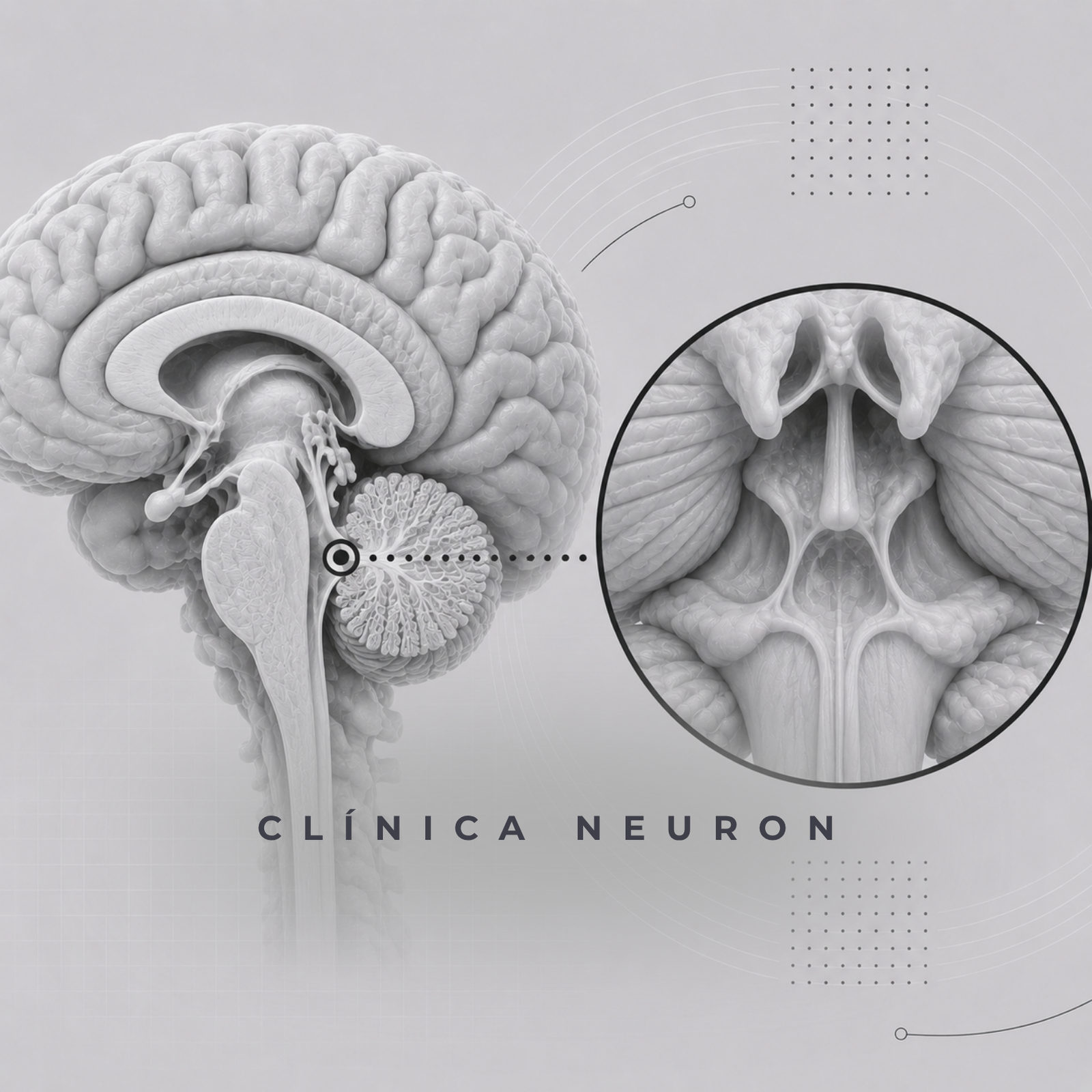
Results The IV ventricle in a midline sagittal cut shows a tentshaped cavity with its roofs pointing posteriorly and the floor formed by the pons and the medulla.
The superior roof is formed by the superior cerebellar peduncles laterally and the superior medullary velum on the midline. The inferior roof is formed by the tela choroidea, the velum medullary inferior, and the nodule.
The floor of the IV ventricle has a rhomboid shape. The rostral two thirds are related to the pons, and the caudal one third is posterior to the medulla. The median sulcus divides the floor in symmetrical halves. The sulcus limitans runs laterally to the median sulcus, and the area between the two sulci is called the median eminence.
The median eminence contains rounded
prominence related to the cranial nucleus of facial, hypoglossal, and vagal nerves. The lateral recesses are extensions of the IV ventricle that opens into the cerebellopontine cistern. The cerebellomedullary fissure is a space between the cerebellum